Additional Diagnostics
Abdominal Ultrasound:
Findings: The spleen appeared within normal limits as did the kidneys. There was echogenic material within the urinary bladder that swirled when the animal’s position was shifted. Both adrenals appeared within normal limits. A large, well circumscribed, well encapsulated mass lesion was noted in the left ventral cranial abdomen. This mass came off the left side of the liver. Most likely the left medial lobe.
Impression: Differentials for echogenic material within the urinary bladder should include white or red cells, cellular debris, proteinaceous material, or mucus. Primary rule-outs for hepatic mass should include neoplasia although granulomatous disease, nodular hyperplasia, and hematoma cannot be completely ruled out. Fine needle aspirate of the junction between the normal liver and mass was done with no immediate complications noted.
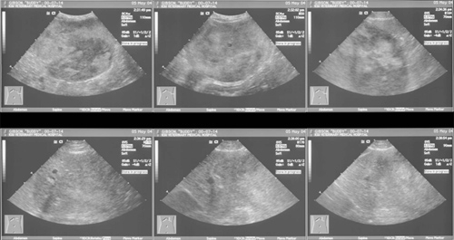
Fig. 5 Ultrasound Images of Hepatic Mass
Fine needle aspirate of liver: Suspect hepatic lipidosis, however aspiration of subcutaneous or peritoneal fat can not be ruled out. The low cellularity of hepatocytes made it difficult to assess the hepatocyte morphology. There were an increased proportion of neutrophils that may indicate inflammation or neutrophilia.
Fine needle aspirate of rectal mass: Inflammatory process with a predominance of eosinophils. The epithelial cells are abnormally arranged and likely represent a dysplastic or hyperplastic process secondary to inflammation. However, an underlying neoplastic process (benign polyp or adenocarcinoma) can not be ruled out.
Three-view thoracic radiographs: No evidence of pulmonary metastatic disease.
Urine culture: E. coli
Abdominal exploratory laparotomy: Removed left medial liver lobe; punch biopsies of liver nodules performed and also culture and sensitivity; jejunal mass removed; rectal mass removed
Histopathology:
Liver mass: hepatocellular carcinoma
Jejunum: jejunal perforation
Rectal masses: rectal adenomatous polyp and rectal epithelial neoplasm
Culture of Liver: no growth
Follow-Up: Administered enrofloxacin and trimethoprim/sulfa antibiotics for urinary tract infection. Improved appetite and attitude.