Interpretation
Radiographic findings:
-
There is a 17 cm soft tissue mass within the left cranioventral abdomen. This mass is causing caudal displacement of the small intestines and spleen and cranial displacement of the gastric axis. The ventral liver margin is incompletely visualized. Within the limited view of the thorax, there is a linear fat opacity in the region of the caudal mediastinal reflection. There is incomplete mineralization of the right 13th rib and absence of the left 13th rib. There are degenerative changes at the coxofemoral joints bilaterally. There is spondylosis deformans at L2-L3 and the lumbosacral junction.
Radiographic Impressions
-
Abdominal soft tissue mass with organs of origin including pedunculated liver, spleen, mesentery, intestine, and pancreas. Differentials for mass include neoplasia, hematoma, abscess/granuloma, and cyst.
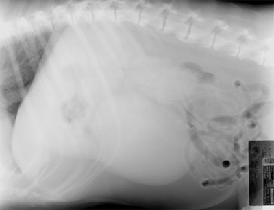
Fig. 3 Lateral Cranial Abdomen
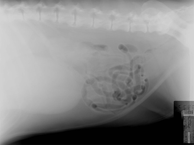
Fig. 4 Lateral Caudal Abdomen