Further Diagnostics
Ultrasound of cranial thorax: Inhomogeneous mass identified in the right cranial thoracic cavity cranial and caudal to the 1st rib. Multiple large vascular structures could be visualized deep to the mass. The mass was immediately adjacent to the 1st rib.
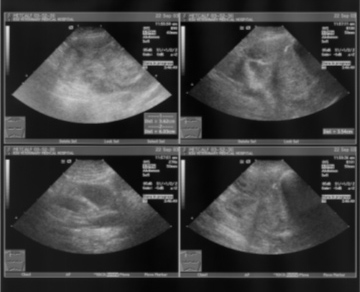
Figure 4. Thoracic ultrasound
CT Findings:
-
There was an ovoid soft tissue mass within the right cranial thoracic cavity which was confluent with the thoracic pleura associated with the 1st rib. There was no evidence of underlying osseous involvement of the 1st rib. There was displacement of the cranial mediastinum and associated vascular structures to the left side adjacent to this mass. The mass was mildly inhomogeneously contrast enhancing which was more pronounced at the periphery of the mass.
CT Impressions:
-
Right cranial thoracic mass most likely originating from the pleura. Rule outs include neoplasia and granulomatous disease. Mass immediately adjacent to the common carotid artery and jugular vein at the thoracic inlet.
Histopathologic diagnosis via true cut biopsy:
-
Hemangiosarcoma