Interpretation
Radiographic Impressions:
Excretory Urography:
A urethral catheter was placed and 10ml of urine was aspirated. 40ml of nitrous oxide was administered via the urethral catheter. 8ml of Hypaque was administered via a left cephalic catheter. The nephrogenic phase revealed an irregularly shaped small left kidney. Subsequent films revealed an ectopic left ureter which paralleled the urethra in the pelvic canal. The right ureter appeared to enter the urinary bladder caudal to the trigone. Urine was visualized entering the urinary bladder via the right ureter.
Differentials for the irregularly small kidneys include renal infarction or renal fibrosis. Left ectopic ureter and suspect right ectopic ureter.
Surgical Findings:
Bilateral ectopic ureters.

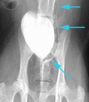
Right lateral oblique view (left up and right down) and ventrodorsal view