Interpretation
Radiographic findings:
-
Pelvis: There is adequate coverage of the femoral heads by the dorsal acetabular rims bilaterally. The coxofemoral joints are congruent bilaterally. There is minimal osteophyte formation at the left cranial acetabular rim. There are multifocal poorly circumscribed regions of increased medullary opacity within the proximal femoral diaphyses bilaterally. The opacities are centered around the nutrient foramina. There are poorly defined radiopaque linear opacities within the mid femoral diaphyses bilaterally.
-
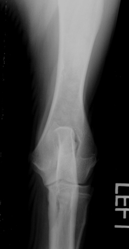
Left Stifle: The previously described poorly defined linear radiopacity wihin the mid femoral diahysis is again present on the lateral view. There is minimal osteophyte formation on the apex of the patella.
-
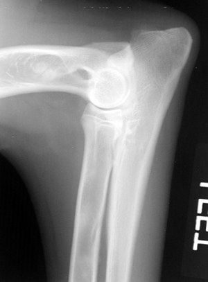
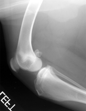
Left Elbow: There are multifocal poorly circumscribed nodular medullary opacities within the distal humeral metaphysis and within the visible portion of the radial medullary cavity on the lateral view.
Radiographic Impressions:
-
Minimal degenerative joint disease of the left coxofemoral and stifle joints. Panosteitis of the femurs bilaterally, left humerus and left radius. The poorly defined linear mid diphyseal opacities within both femurs may be a manifestation of panosteitis or may be incidental growth arrest lines.
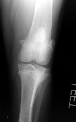
Craniocaudal Left Stifle
Craniocaudal Left Elbow
Lateral Left Elbow
Lateral Left Stifle