Interpretation
Radiographic findings:
-
Left stifle: There is increased synovial mass marked by cranial displacement of the infrapatellar fat pad and bulging of the caudal joint pouch. There is irregular lucency of the subchondral bone of the left lateral femoral condyle. There is flattening of the subchondral bone of the left lateral femoral condyle. There is mild sclerosis surrounding the lucent area of the left femoral condyle. There is irregular new bone formation at the medial and lateral condyles of the tibia and at the base and apex of the patella. There is an irregular mineral opacity (approximately 5 mm X 5 mm) within the caudal joint space.
Radiographic Impressions:
-
Lucency and flattening of the subchondral bone at the lateral femoral condyle are consistent with osteochondrosis of the left stifle. Increased synovial mass is most consistent with osteoarthrosis, likely secondary to osteochondrosis. The mineral opacity in the caudal joint space likely represents a mineralized cartilage fragment (joint mouse). Recommend radiographs of the right stifle for comparison.
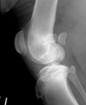
Fig. 3 Left stifle. Lateral oblique view.
What are your radiographic findings? What is the most likely diagnosis? What is your recommended treatment?