Case Follow-Up
Further Diagnostics:
-
Abdominal ultrasound findings and impressions: inhomogeneous eccentrically located intestinal mass in the right caudal abdominal cavity, multiple enlarged hypoechoic lymph nodes with central portions of increased echogenicity at the root of the mesentery/ rule outs for the intestinal mass include neoplasia with lesser consideration given to granulomatous infiltration, rule outs for mesenteric lymphadenopathy include neoplasia and reactive lymph nodes.
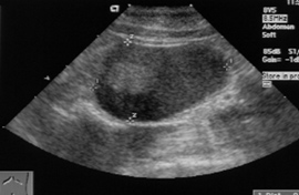
Figure 1: Mesenteric Lymph Node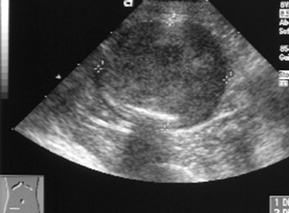
Figure 2: Intestinal Mass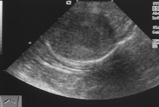
Figure 3: Intestinal Mass
-
Fine needle aspirates of intestinal mass and lymph nodes (ultrasound guided): nondiagnostic
-
Intestinal resection and anastomosis performed, submitted surgical biopsies of intestinal mass and lymph nodes: malignant neoplasm, poorly differentiated mast cell tumor
Follow-up:
-
Owners elected chemotherapy and the cat has an improved appetite and gained weight.