Warren Beard, DVM, MS, Diplomate ACVS
What is the guttural pouch?
A.
B.
C.
D.
E.
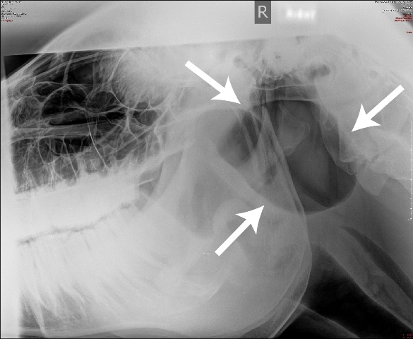
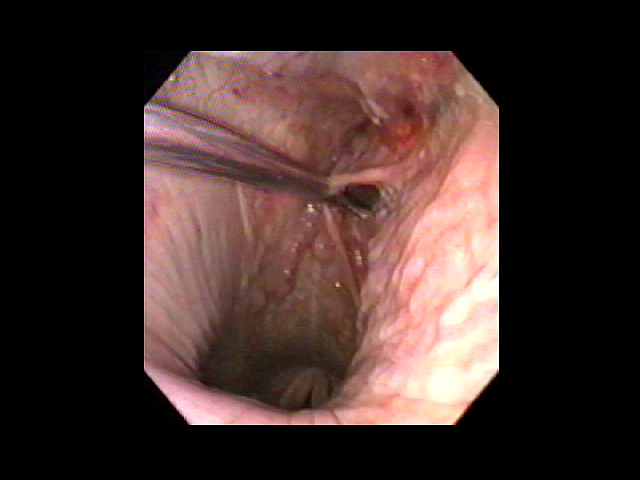
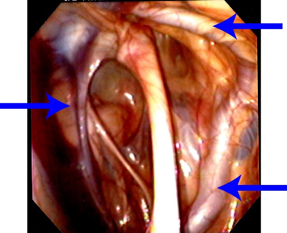
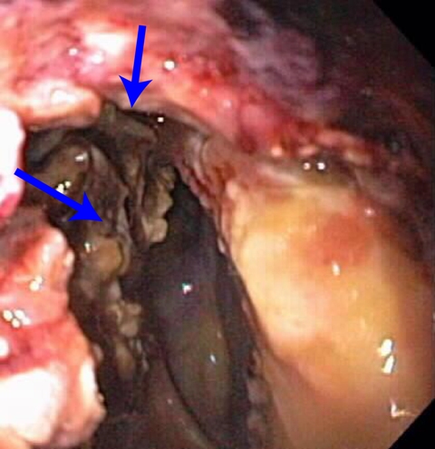
<tr> <td> <p>F.</p> <p> <img src="/images/vhc/timely-topics/image7.gif" alt="guttural pouch radiograph" width="256" height="192"> </p> </td> </tr> <p>There is a small tube that connects the inner ear to the pharynx or throat called the Eustachian tube. It allows the pressure between the inner ear and the throat to equilibrate. It is the reason you have to swallow on airplanes when you are ascending and descending. Horses are unique in that they have a diverticulum off of the Eustachian tube called the guttural pouch. It is an air filled structure about the size of a baseball just below the horse's ear. (Image A)</p> <div>Because the guttural pouch is positioned at the base of the skull, some very important nerves and arteries course through it. The nerves that control swallowing, hearing, balance, and facial expression course through the guttural pouch. The three largest arteries supplying blood to the head, the internal carotid, the external carotid, and the maxillary arteries all course through the guttural pouch.</div> <div>Endoscopic view of the nasopharynx with a catheter holing open the opening to the guttural pouch. (Image B)<br> <br>Endoscopic view of the guttural pouch demonstrating the internal carotid artery, the external carotid artery, and the maxillary artery. (Image C)</div> <div></div> <h4> <strong>Why a guttural pouch?</strong> </h4> <div>The purpose of the guttural pouch is unknown. It is just the way horses are made. The significance of the guttural pouch lies in the fact that it is a hollow air filled structure that communicates with the throat every time the horse swallows. This communication with the throat allows the important nerves and arteries to come into contact with potential pathogens in the form of bacteria and fungi.</div> <div></div> <h4> <strong>What is guttural pouch mycosis?</strong> </h4> <div>Guttural pouch mycosis is a fungal infection in the guttural pouch caused by a common fungi that most all horses carry. The fungus has an affinity for growing on the surface of the guttural pouch overlying the nerves and arteries. The fungus growing on nerves causes paralysis of affected nerve while the fungal plaques on the arteries will erode through the arterial wall causing life threatening or fatal hemorrhage. It is not known why certain horses are affected by a fungus that is a normal contaminant of all horses.</div> <div>Fungal plaques growing on the surface of the guttural pouch lining and obscuring the normal anatomy. (Image D)</div> <div></div> <h4> <strong>Clinical signs</strong> </h4> <div>Horses affected by guttural pouch mycosis typically present for acute onset of voluminous hemorrhage from the nares. Many horses will exsanguinate on the first episode.</div> <div></div> <h4> <strong>Treatment</strong> </h4> <div>Emergency treatment in the form of intravenous fluids and blood transfusion is necessary to save the horses life. Occluding the affected arteries is necessary to stop the hemorrhage. The preferred method of occluding the arteries is by a method called coil embolization. The carotid artery is catheterized and coils resembling miniature pipe cleaners are placed in each of the arteries under fluoroscopic guidance. Prognosis for horses that undergo coil embolization is good and most are able to return to their previous level of activity. <br> <br>Video of a coil being placed in the internal carotid artery under fluoroscopic guidance. The linear black structure is the carotid artery highlighted by intraarterial injection of contrast medium. (Image E)<br> <br>Radiograph of the horses guttural pouch demonstrating coils placed in the internal carotid artery, the external carotid artery, and the maxillary artery. (Image F)</div> <p></p>